This week we discuss Lisfranc injuries with a focus on a diagnostic pathway and management.

Audio is streamed directly from the publisher (media.blubrry.com) as published in their RSS feed. Play Podcasts does not host this file. Rights-holders can request removal through the copyright & takedown page.
Show Notes

Show Notes
Take Home Points
- A Lisfranc injury is a midfoot injury that results in displacement of one or more of the metatarsal bones from tarsus.
- XR will show widening of the space between the 1st and 2nd metatarsals. Getting contralateral XR may help you identify this.
- Even if you don’t see that widening on the XR, the patient could still have a Lisfranc injury. If they cannot walk due to pain, get a weight bearing XR or CT scan to look further.
- Once the injury is identified, the patient must be strict non-weightbearing. Place them in a posterior splint and get orthopedics involved either in the ED or for prompt follow up as the patient will probably need surgery.
Foot Bones (Google Images)
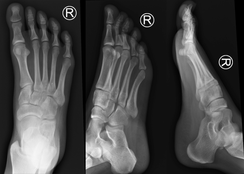
Normal Foot X-ray Series (Case courtesy of Dr Andrew Dixon, Radiopaedia.org. From the case rID: 36688)
Lisfranc Injury AP X-ray (Radiopaedia Image #1: Case courtesy of Dr Alexandra Stanislavsky, Radiopaedia.org. From the case rID: 10919)
Divergent Lisfranc Injury
Read More
LITFL: Eponymous Fractures
Radiopaedia: Lisfranc Injury
Core EM: Compartment Syndrome
Read More